
A number of research and medical projects depend on our technology

DIVA presents 3D imaging data it should be viewed

Stem Cell Dynamics

Spatial dynamics of stem cells during development, Institut Pasteur


The team of Pauline Spéder at the Institut Pasteur is using DIVA to comprehend the spatial organization of neuronal stem cells in the drosophila larva. It has notably been used to visually comprehend and visualize the exchange of cytoplasmic materials between fused cells in the brain


Immune response to pathogens

Dynamics of Immune Responses, Institut Pasteur

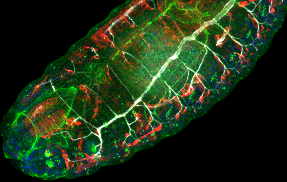
The team of Philippe Bousso at the Institut Pasteur studies the spatiotemporal characteristics of the immune response to pathogens in zebra fish. Using two-photon confocal imaging data, DIVA has been used to understand the how leukocytes find and identify their targets

Institut Pasteur

Image Analysis Hub


DIVA is installed in the Image Analysis Hub at the Institut Pasteur, where researchers have privileged access to view and quantify their volumetric images


Pre-operative planning

Institut Curie Hospital

In collaboration with the Institut Curie hospital, DIVA is being used as a preoperative tool to enhance the perception of breast cancer in MRIs for surgeons and radiologists alike

Institut Curie

Available to the entire Institut Curie


DIVA is currently installed in the Institut Curie Nikon Imaging Center where it is at the disposition of all researchers in the Institut Curie research center

